Latest news


Anti-GAD65 antibodies: Clinical Evidence and Practical Advantages of RIA vs CLIA tests
Understanding Anti-GAD65 Antibodies and Their Clinical Significance
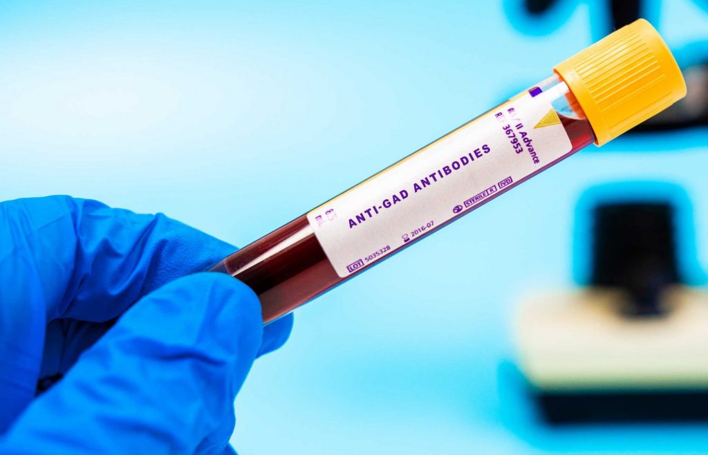
Anti-glutamic acid decarboxylase antibodies (anti-GAD65) are among the most important biomarkers in autoimmune diseases. They play a dual diagnostic role:
- at low to moderate titers, they help identify autoimmune diabetes, particularly type 1 diabetes;
- at very high titers, they strongly suggest neurological autoimmune conditions such as stiff-person syndrome, autoimmune encephalitis, cerebellar ataxia, and some forms of epilepsy.
These antibodies can reach concentrations 100–500 times higher in neurological disease compared with diabetes, making quantitative accuracy crucial.
A recently published scientific study evaluated the clinical performance of anti-GAD65 determination of a commercial chemiluminescent immunoassay (CLIA) and compared it with a traditional radioimmunoassay (RIA from Medipan)1. Since automated CLIA are increasingly adopted in many labs due to their intrinsic practical advantages, the authors studied 90 patients (54 with autoimmune diabetes and 36 with neurological GAD-spectrum disorders). The findings highlight significant challenges and reveal that neither method is immune to analytical obstacles, especially at extremely high titers. These analytical considerations are increasingly relevant as type 1 diabetes care evolves toward earlier detection and prevention strategies. In this emerging scenario, accurate measurement of anti-GAD65 antibodies is critical for distinguishing autoimmune diabetes from neurological GAD65-spectrum disorders, particularly when antibody titers reach extreme levels.
What the Study Shows: Key Clinical and Analytical Conclusions
1. CLIA consistently overestimates titers compared with RIA
The CLIA produced values approximately five times higher than RIA on average, demonstrating a significant proportional bias, especially at higher concentrations. Agreement was excellent in diabetic patients but substantially weaker in neurological cases, where titers were highest. This indicates that CLIA becomes progressively less linear as concentrations increase.
2. Identification of a diagnostic cutoff to differentiate neurological from diabetic patients
Based on ROC analysis, the authors identified an optimal threshold, achieving 94–96% accuracy in distinguishing neurological GAD65 autoimmunity from type 1 diabetes. This distinction is essential because low-level anti-GAD65 positivity is common in diabetes but rarely clinically meaningful in neurology.
3. Both CLIA and RIA can experience hook effect—but CLIA does so silently
A key finding concerns the hook effect. The CLIA showed several neurological samples with unexpectedly low undiluted results that, after dilution, revealed true, very high titers. This is clinically dangerous because values in the mid-range can appear to be normal. RIA also exhibited hook effect in neurological samples—100% incidence when run undiluted—but its behavior was predictable: once diluted, it returned to full linearity and accurately revealed the true titers. This contrast sets the stage for understanding the practical advantages of RIA.
Why RIA Still Matters: Practical and Analytical Advantages in High-Titer Anti-GAD65 Testing
1. Predictable and transparent behavior
Although RIA has an intrinsic upper limit due to tracer precipitation, this saturation results in stable, high plateau values that are readily recognizable as analytical saturation rather than deceptively low measurements. In contrast, CLIA may generate falsely low values within a seemingly normal range, making underestimation more clinically dangerous. This makes RIA particularly trustworthy for neurological samples, where extremely high concentrations are expected.
2. Full recovery of linearity after dilution
Unlike CLIA—which shows progressive non-linearity and dispersion before reaching extreme titers—RIA maintains stable proportionality once concentration falls within the measurable range. This ensures that even high titers can be quantified accurately.
3. A dependable reference method for assay calibration
The study relies on RIA to evaluate the CLIA, establish cutoffs, and quantify proportional bias. RIA serves as the anchor method against which new assays are validated, reflecting decades of accumulated clinical experience.
4. Reduced risk of “silent” misclassification
CLIA may underestimate titers even at moderate ranges, but RIA’s failure mode is obvious and correctable. For neurological syndromes—where diagnosis hinges on recognizing extremely high titers—this transparency is a clear advantage.
Conclusion

In complex autoimmune diagnostics, not all assays fail in the same way. The article demonstrates that while no method is impeccable at extremely high anti-GAD65 concentrations, RIA offers predictability, reliable post-dilution linearity, and a trusted reference framework.
Understanding how RIA and CLIA behave at extreme titers of anti-GAD65 is essential to avoid silent misclassification. For laboratories dealing with neurological autoimmunity and early risk stratification in type 1 diabetes, a reference-grade method such as the Medipan RIA remains crucial.
Product used in the study
| RIA | 2070, 2071 – CentAK® anti-GAD65 M | Quantitative determination of antibodies against Glutamic Acid Decarboxylase (GAD65) |
Type 1 Diabetes Related products
| ELISA | 3507 – Medizym® anti-GAD M | Quantitative determination of antibodies against Glutamic Acid Decarboxylase (GAD65) |
| ELISA | 3506 – Medizym® anti-IA2 M | Quantitative determination of antibodies against Protein Tyrosine Phosphatase (IA2) |
| RIA | 2050, 2150 – CentAK® anti-IA2 M |
| ELISA | 3806 – Medizym® IAA | Quantitative determination of antibodies against Protein Tyrosine Phosphatase (IA2) |
| RIA | 2035 – CentAK® IAA M |
| IFA for standard microscope | 85848 – ICA IFA | Determination of IgG antibodies against islet cells (ICA) |
| IFA for akiron®NEO | 4129 – AKLIDES® ICA |