News

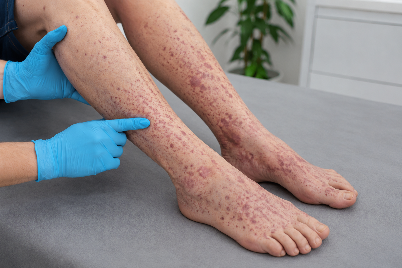
A healthcare provider wearing blue medical gloves examines a patient's lower legs and feet, which are densely covered in a reddish-purple rash characteristic of vasculitis or purpura, while the patient rests on a grey examination table.
23.07.2026

ANCA-associated vasculitis: the 2022 ACR/EULAR classification criteria and the role of IVD antibody testing

ANCA antibody testing plays a central role in the 2022 American College of Rheumatology/European Alliance of Associations for Rheumatology (ACR/EULAR) classification criteria for ANCA-associated vasculitis (AAV). A recent study evaluating these criteria in children provides a useful entry point into the classification of AAV. Although the criteria were developed from adult cohorts, the pediatric analysis […]
More details
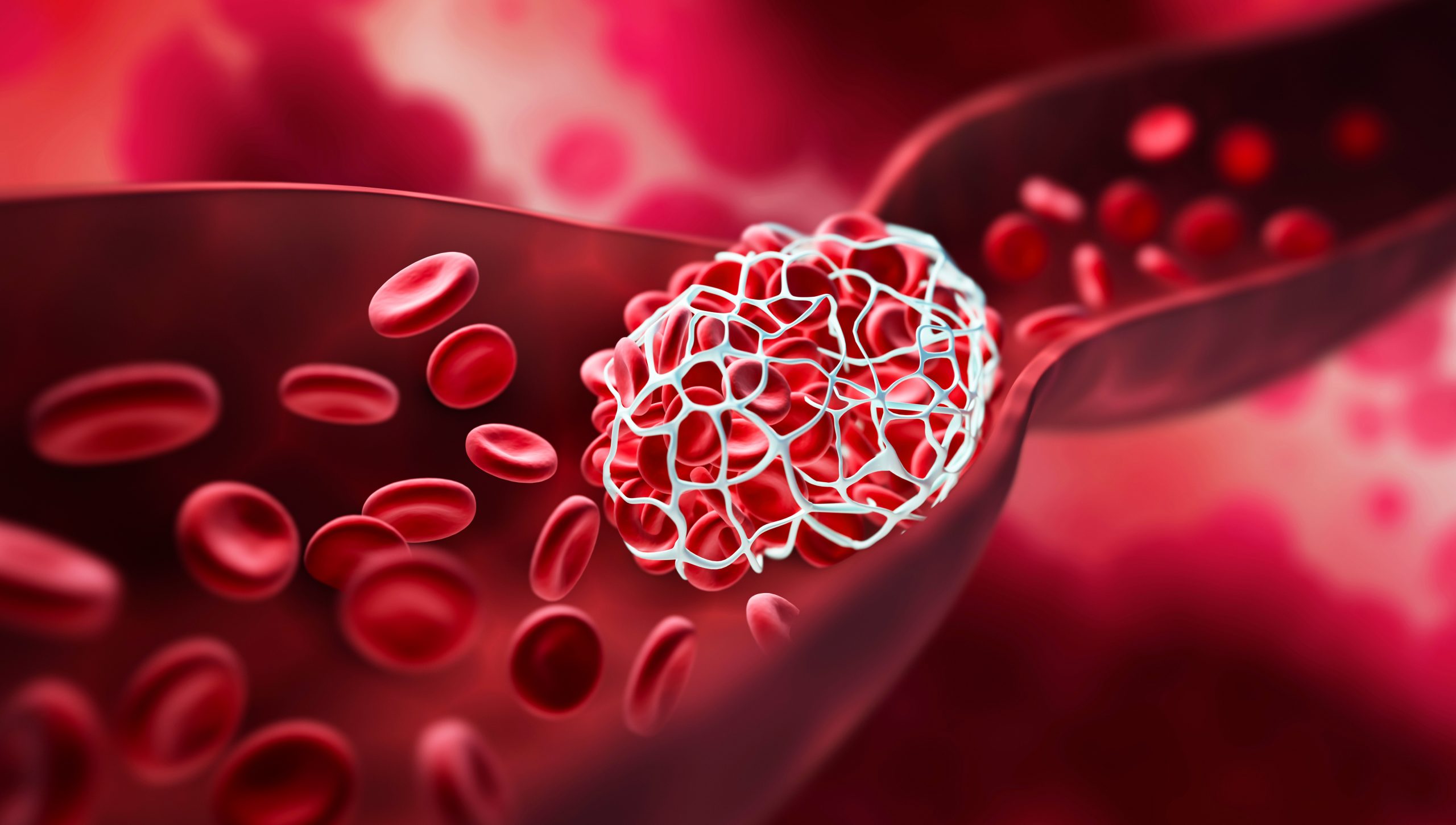
The image is a medical illustration of a blood clot forming inside a blood vessel. It shows red blood cells flowing around a large, white, mesh-like structure.
18.06.2026

Anti-phospholipid Antibodies (aPL) as Prognostic Markers

Anti-phospholipid antibody (aPL) testing is used in the evaluation of suspected antiphospholipid syndrome (APS). aPL may also be detected in patients with systemic lupus erythematosus (SLE) and can inform the assessment of APS-related thrombotic or obstetric risk when interpreted together with established clinical criteria. A recent 10-year cohort study published in The American Journal of […]
More details
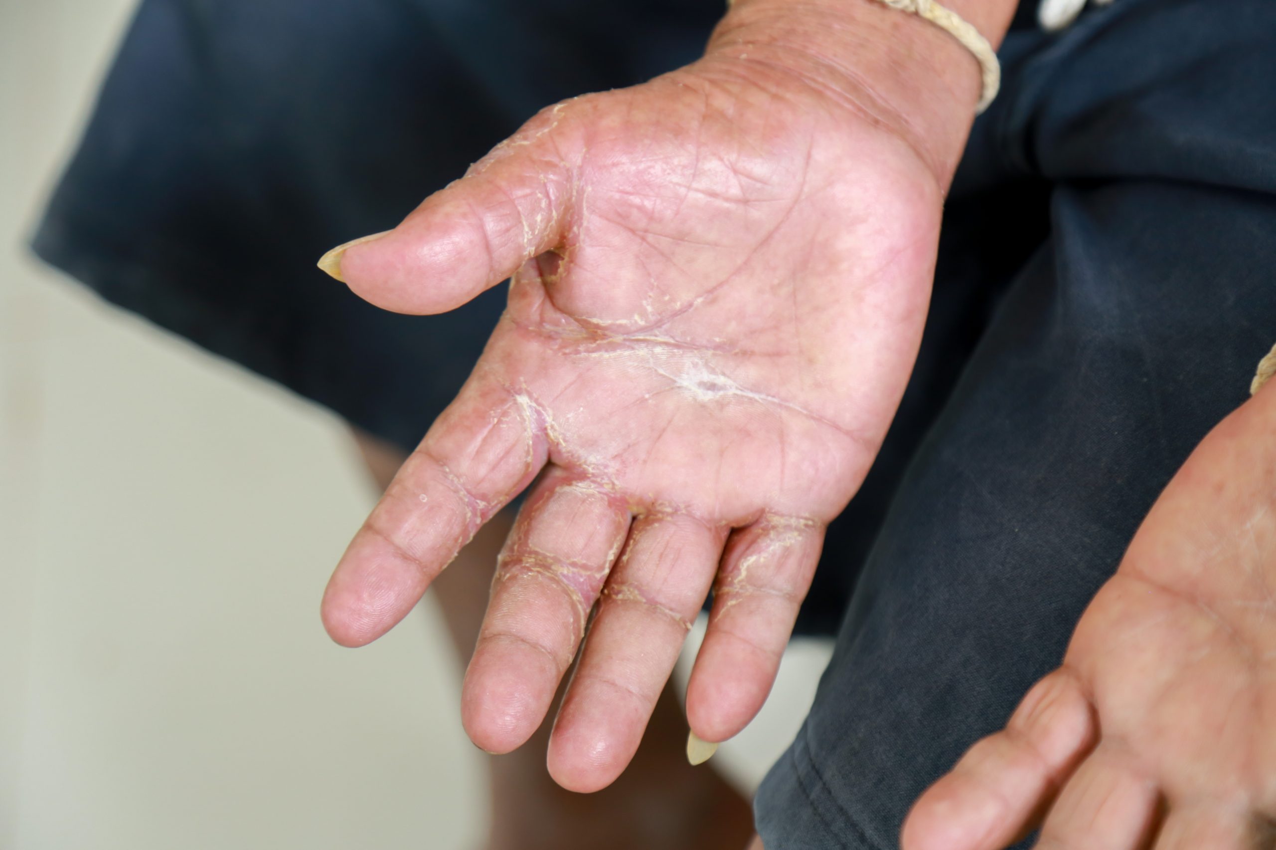
The image shows the open palm of a person with visible signs of scleroderma. The skin on the hand is thickened, taut, and appears shiny. Deep, cracked lines are noticeable at the joints and across the palm, indicating a hardening and stiffening of the skin. The skin looks dry and scaly, and the fingers appear swollen.
01.06.2026

Understanding Scleroderma and Its Diagnostic Tests

June is recognized as Scleroderma Awareness Month to honor those affected by the disease. The month ends with World Scleroderma Day on June 29. This date commemorates Swiss painter Paul Klee, who died of systemic sclerosis in 19401. Scleroderma, or systemic sclerosis, is a rare autoimmune disease. It causes hardening and tightening of the skin […]
More details
A modern laboratory setting featuring two scientists in the background wearing white lab coats and looking at a tablet. In the foreground sits the "akiron® NEO" analyzer with an integrated screen displaying an immunofluorescence image (bright green dots on a black background). Pipettes, a barcode scanner, and a microscope are also visible on the lab bench.
01.06.2026

AI-Based ANA HEp-2 IFA Interpretation: Clinical Validation in Routine Diagnostics

Antinuclear antibody (ANA) testing by indirect immunofluorescence assay (IFA) on HEp-2 cells is widely used as a reference screening method for systemic autoimmune rheumatic diseases (SARD), including systemic lupus erythematosus, systemic sclerosis, and Sjögren’s syndrome. However, manual interpretation of ANA HEp-2 IFA patterns is time-consuming, operator-dependent and subject to inter-observer variability. A recent study published […]
More details
Anfrage erfolgreich Gemini hat gesagt This image features a medical doctor, explaining human anatomy using a detailed anatomical model of the liver and gallbladder.
31.05.2026

Serological Biomarkers in Primary Sclerosing Cholangitis (PSC): Focus on anti-GP2 IgA

Primary sclerosing cholangitis (PSC) is a chronic, progressive inflammatory disease affecting the bile ducts and often leading to fibrosis, cirrhosis, and eventually liver failure. PSC also carries an increased risk of hepatobiliary malignancies, particularly cholangiocarcinoma. Despite significant research efforts, PSC remains difficult to diagnose and manage because its pathogenesis is not fully understood and reliable […]
More details
Two female scientists work in a modern equipped computer laboratory analyzing blood samples and genetic materials using special machines in a modern laboratory.
10.03.2026

Variability in Diagnostic Test Results

Understanding Normal Analytical Variation In diagnostic testing, it is not uncommon to observe small differences when the same sample is measured repeatedly. Such variations are expected and reflect the inherent characteristics of the assay, even under well-controlled laboratory conditions. Diagnostic assays rely on complex biological, chemical, and technical processes. Each step introduces a degree of […]
More details
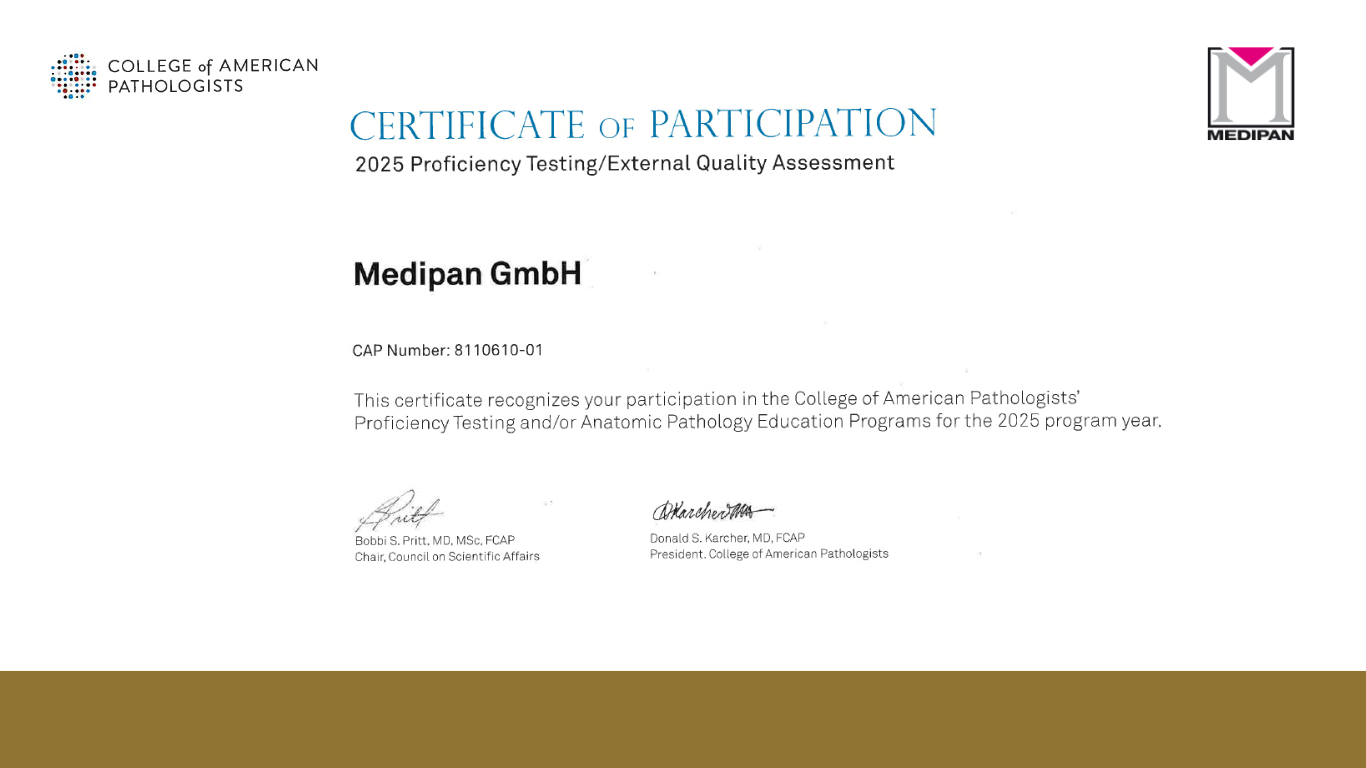
A "Certificate of Participation" from the College of American Pathologists (CAP) issued to Medipan GmbH. The certificate recognizes participation in proficiency testing or anatomic pathology education programs for the 2025 program year. It includes CAP number 8110610-01 and signatures from Bobbi S. Pritt and Donald S. Karcher. The Medipan logo is displayed in the top right corner.
20.02.2026

Celebrating 8 Years of Excellence in the CAP Antinuclear Antibody (ANA) Survey!

Celebrating 8 Years of Excellence in the CAP Antinuclear Antibody (ANA) Survey! We are proud to announce an important milestone in our quality journey: Eight consecutive years of successful participation in the Antinuclear Antibody (ANA) Survey organized by the College of American Pathologists (CAP). Eight Years of Commitment to Quality For eight years, our laboratory […]
More details
A laboratory professional wearing protective gloves holds a 96-well microtiter plate featuring the Anti-Factor H ELISA assay. The wells of the plate contain various shades of blue, indicating positive test results. The image includes the Medipan & GA Generic Assays logo in the upper right corner.
02.02.2026

Anti–Factor H Autoantibodies and ELISA Testing: Optimizing Diagnosis in Atypical Hemolytic Uremic Syndrome (aHUS)

Atypical hemolytic uremic syndrome (aHUS), also known as complement-mediated thrombotic microangiopathy (CM-TMA), is a rare but life-threatening condition caused by dysregulation of the alternative complement pathway. Among the known etiologies, autoantibodies against complement factor H (anti-factor H) play a critical role, accounting for approximately 10% of cases. Accurate laboratory detection of these antibodies is essential, […]
More details
3D illustration of Vibrio cholerae bacteria in the lumen of the small intestine. The image highlights the characteristic curved, comma-shaped morphology and the single polar flagellum of the bacteria.
12.01.2026

Cholera and Guillain–Barré Syndrome: When Epidemics Trigger Autoimmune Neuropathy

In regions affected by recurrent cholera outbreaks, overlapping infectious diseases can obscure the diagnosis of post-infectious autoimmune conditions. We recently collaborated in the publication describing a pediatric case from Yemen that illustrates how Guillain–Barré Syndrome (GBS) can emerge during a cholera outbreak, highlighting the critical role of Campylobacter jejuni infection and anti-ganglioside antibody testing 1. […]
More details
The image shows a smiling, blonde woman in a laboratory. She is wearing a white lab coat and safety goggles. She holds up a test tube containing a blue liquid with her gloved hand. In the blurred background, another person in a lab coat is also visible. The setting appears to be a modern lab with shelves and equipment
01.01.2026

Product Catalog 2026

Discover the 2026 Product Catalog – The Latest in Autoimmune Diagnostics Medipan GmbH and GA Generic Assays GmbH proudly present the 2026 Product Catalog, showcasing our expanded portfolio of diagnostic solutions for autoimmune diseases. This edition highlights the latest innovations in immunodiagnostics and equips healthcare professionals, researchers, and laboratory partners with tools to advance diagnostics […]
More details
Contact us