
Latest news









AI-Based ANA HEp-2 IFA Interpretation: Clinical Validation in Routine Diagnostics
Antinuclear antibody (ANA) testing by indirect immunofluorescence assay (IFA) on HEp-2 cells is widely used as a reference screening method for systemic autoimmune rheumatic diseases (SARD), including systemic lupus erythematosus, systemic sclerosis, and Sjögren’s syndrome1. However, manual interpretation of ANA HEp-2 IFA patterns is time-consuming, operator-dependent and subject to inter-observer variability2.
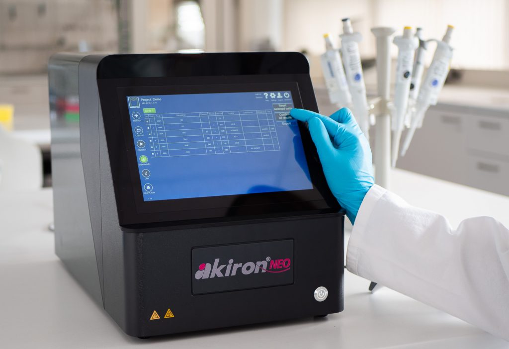
A recent study published in Clinica Chimica Acta compared expert visual interpretation of ANA HEp-2 IFA with AI-assisted image analysis using the evaluated akiron® NEO configuration under routine laboratory conditions3. The investigation included 2,671 consecutive serum samples analyzed in a routine diagnostic laboratory and compared expert visual reading with automated image interpretation using the akiron® NEO. This analysis is based on the solid results already emerging from a pilot study that had previously highlighted the potential of artificial intelligence in supporting ANA diagnostics4.
Real-World Performance of Automated ANA Diagnostics
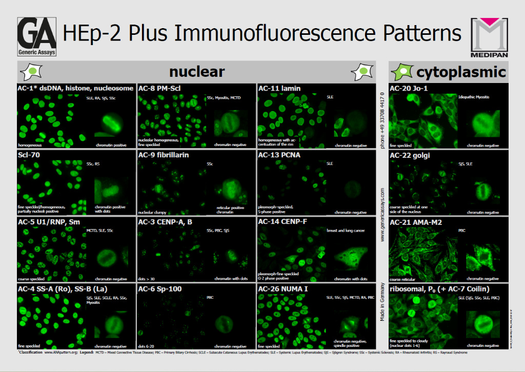
The study assessed ANA positivity/negativity discrimination, titer assignment, and fluorescence pattern recognition according to the International Consensus on ANA Patterns (ICAP) at the competent level.
A good concordance was found for positive/negative ANA classification (κ 0.616–0.699). Agreement improved further when weakly positive samples were excluded.
Pattern recognition performance was similarly robust. Nuclear patterns reached kappa values up to 0.650 in clinically significant titers (≥1:320), while mitotic pattern identification achieved even higher agreement (up to κ = 0.736). Cytoplasmic patterns showed slightly lower concordance, consistent with the inherent variability described in ANA diagnostics literature5.
Under the conditions of this study, agreement between the evaluated AI-assisted analysis and expert interpretation was within the range of inter-observer agreement reported between experienced immunologists. This finding indicates that variability in ANA interpretation reflects intrinsic methodological complexity rather than technological limitation. These findings apply to the configuration evaluated in this study and should not be generalized to other assay configurations or laboratory settings.
The automated system showed a slightly lower overall positivity rate than one human observer. This finding suggests a more conservative classification of borderline cases. In routine laboratories, especially outside specialized rheumatology settings, this approach may help reduce unnecessary follow-up testing after weakly positive results.
Deep Learning Architecture for ANA Pattern Recognition
The akiron® NEO platform applies hierarchical convolutional neural networks (CNNs) trained for specific analytical tasks, including cell cycle phase detection, nuclear and cytoplasmic pattern recognition, and mitotic analysis. DAPI counterstaining supports accurate metaphase identification, which is essential for ICAP-compliant classification. The system integrates these outputs to generate standardized ANA HEp-2 IFA interpretations aligned with ICAP nomenclature. The performance described in this article refers to the software version and system configuration evaluated in the cited study.
Clinical Correlation
In a subset of samples with reflex immunoblot testing, AI-based pattern recognition demonstrated expected serological associations. For example, all AC-3 centromere patterns corresponded to anti-CENP-B positivity, while speckled patterns showed associations with anti-Ro52, SS-A, SS-B, Sm and U1-nRNP antibodies. These findings support the clinical validity of AI-driven ANA classification.
Conclusion
In the cited study, the akiron® NEO configuration achieved agreement with expert interpretation comparable to the reported inter-observer agreement among experienced immunologists under the study conditions. Automated image analysis may support laboratory workflow standardization and reproducibility but does not replace expert review or clinical correlation.
The reported findings suggest that AI-assisted image analysis with akiron® NEO may support standardized ANA HEp-2 IFA interpretation in routine laboratory workflows. Performance depends on the evaluated assay configuration, software version, laboratory procedures, and implementation.
Study Scope and Limitations
The reported results are based on a single routine diagnostic laboratory using the akiron® NEO hardware and software configuration evaluated in the cited study. The study included 2,671 consecutive serum samples analyzed under routine laboratory conditions. Performance may vary depending on assay configuration, software version, laboratory workflow, sample characteristics, and user implementation. Automated image interpretation supports, but does not replace, expert review and clinical correlation.
Product used in the study
| 4450 – akiron® NEO | IFA analyzer with AI-based software used for automatic ANA, ANCA, nDNA and CytoBead® pattern recognition and intensity/titer assessment |
| 4063, 4065 – AKLIDES® ANA plus | HEp-2 cells based IFA for the determination of IgG antibodies against nuclear and cytoplasmic antigens (ANA) |
Discover the full range of the immunofluorescence products
References
- Bossuyt et al., (2020) Understanding and interpreting antinuclear antibody tests in systemic rheumatic diseases ↩︎
- Rigon et al., (2017) The inter-observer reading variability in anti-nuclear antibodies indirect (ANA) immunofluorescence test: a multicenter evaluation and a review of the literature ↩︎
- Schmidt et al., (2025) Comparison of manual with AI-aided interpretation of ANA HEp-2 IIF assay patterns in a clinical diagnostics lab ↩︎
- Mayr et al., (2025) Pilot Study of AI-Assisted ANA Immunofluorescence Reading—Comparison with Classical Visual Interpretation ↩︎
- Rigon et al., (2017) The inter-observer reading variability in anti-nuclear antibodies indirect (ANA) immunofluorescence test: a multicenter evaluation and a review of the literature ↩︎