4319 – AKLIDES® Immunology
Highlights
• Automated IFA analyzer for high sample throughput
• Digital imaging of processed IFA slides
• ANA / ANCA pattern recognition and intensity evaluation
• ANA / ANCA end-titer determination from only one standard sample dilution
• CLIFT for determination of antibodies against dsDNA
• Results in 35 seconds*
• Imaging of tissue sections (e.g. EmA, …)
• Quantification of antibody activities in U/mL or IU/mL using CytoBead® technology
• Archiving of images and results
• Quality assured data management
• Export of all relevant results in pdf- or xls-file format
• LIS Connectivity
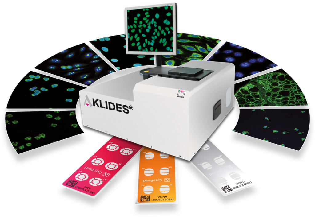
The AKLIDES® system is the world’s first automated IFA analyzer for standardized, digital imaging of processed IFA slides. The AKLIDES® system is used to support the diagnosis of autoimmune diseases. It allows the analysis of up to five slides or 60 samples and is designed for a high sample throughput.
The user-friendly AKLIDES® software enables objective ANA / ANCA pattern recognition in approx. 35 seconds per sample (ANA / ANCA / CLIFT). The simultaneous recording of the fluorescence intensity allows the determination of the end-titer from just one standard sample dilution, making the analysis of complex and cost-intensive dilution series obsolete.
Validated protocols and AKLIDES® assay files support the standardized evaluation of numerous immunofluorescence analyzes for the determination of antibodies against HEp-2 cells (ANA), granulocytes (ANCA) and Crithidia luciliae (CLIFT, dsDNA), various tissue sections and against specific antigens using the CytoBead® technology. The results are clearly presented in well-arranged reports.
The powerful AKLIDES® system is easy and intuitive to use and indispensable for all diagnostic routine services in the fields of rheumatology and gastroenterology.
* ANA / ANCA / CLIFT
Product Specifications
| Title | AKLIDES® |
| Product Code | 4319 |
| Dimensions / Weight | 65 cm x 67 cm x 32 cm (L x W x H) / 78 kg |
| Indication | Autoimmune Diseases |
| Description | Automated System for Standardized Immunofluorescence Imaging |
| Optics | LED (4 wavelengths) |
| Objectives | 2 different Objectives (10x, 40x) |
| Filters | blue / green / orange / red |
| Capacity | 5 Slides |
| Analysis | Cell and Tissue Imaging, Cell Pattern Recognition, Intensity Evaluation, End-Point Titer Determination |
| Parameters | ANA, ANCA, EmA, nDNA, CytoBead®, Tissue Sections |
| Report | Overall and Single Report, Documentation, Archival Storage |
| Connectivity | Pipettor and bidirectional to LIS |
Free downloads
Flyer [REF 4319][eng] Flyer [REF 4319][deu] Flyer [CytoBead Technology][eng] Flyer [Celiac Overview][eng]Restricted downloads - Password required
Current version of the instructions for use. The respective valid version for processing the test can be found in the product packaging.